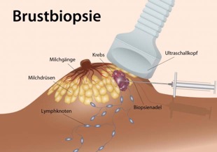
Removal of tissue samples from the breast/ mammary graft biopsy
For breast cancer screening, regular breast self-examination is recommended, as well as routine examination as part of the annual check-up. But what happens next if a woman has felt a lump in her breast or an abnormality is found in the early detection examination?

The next step is then usually an imaging examination (mammography and or mammary sonography/breast ultrasound). Especially for women under 40 with comparatively dense breast tissue, ultrasound is the imaging examination of choice and can be performed in our practice. This ultrasound as well as mammography and MRI (magnetic resonance imaging) allow a very precise classification of the abnormalities found. However, if there are unclear changes in these examinations, it may be necessary to take a tissue sample. The tissue obtained can then be examined under a microscope and a reliable diagnosis can be made. With this procedure, most abnormal or unclear breast findings can now be clarified minimally invasively and without surgery.
In our practice, we perform ultrasound-guided punch biopsy to clarify most palpable changes in the breast or those that can be easily visualized by ultrasound. If a change in the breast can only be visualized by mammography or breast MRI, the tissue sample is taken outside the practice under the control of these procedures.
In punch biopsy, after local anesthesia, a hollow needle is inserted ultrasound-guided in front of the finding and then automatically advances at high speed into the nodule or area to be examined to remove a tissue sample. Due to the local anesthetic and the high speed at which the sample is taken, there is usually no pain. The risk of infection, bleeding and injury to surrounding structures is very low. The probability of tumor cells being carried over is also very small due to appropriate technical measures. In order to obtain a representative tissue sample, at least three punches are taken, thus increasing the significance of the microscopic examination. The result of the histological examination is usually available after 2-3 working days. In the case of a “benign” result, further -particularly- surgical measures can usually be dispensed with. In most cases, the further procedure is then limited to regular follow-up checks. In the case of malignant or uncertain changes, removal or further therapy is planned depending on the findings. After the sample has been taken, a pressure bandage is applied and physical exertion should be avoided for the next 24 hours.
German Cancer Research Center, German Cancer Society
